A hostile takeover: Tumors can program T cells to express PD-L1 and restrain their antitumor responses
During an immune response, T cells play a fundamental role in recognizing and neutralizing abnormal cells and pathogens, such as bacteria, viruses, and tumors. The majority of cells in our body chew up small portions of their proteins and present them on their cell surfaces as antigens. The T cell receptor (TCR) that sits on the surface of T cells acts as a detector that is able to scan thousands of cells and uncover potentially harmful antigens. When T cells encounter these antigens, they mount an attack. There are 2 main types of T cells: (1) CD8 T cells, which actively seek out and destroy harmful cells, and (2) CD4 T cells, which have no killing capacity but assist other white blood cells in their immune responses, including CD8 T cells.
It is important that T cells target only harmful cells – loss of this ability can lead to autoimmune diseases, such as rheumatoid arthritis or multiple sclerosis, where T cells attack healthy cells. Immune checkpoints are inhibitory signals wired into T cells that act as brakes to tamp down T cell responses and are critical in protecting us from developing autoimmune diseases. However, these pathways can also be hijacked by cancer cells to evade immune surveillance. One example of this is the increased expression of a protein called PD-L1 on the surface of cancer cells. PD-L1 binds to the checkpoint surface protein, PD-1 expressed on T cells. PD-1 engagement by PD-L1 suppresses the activity of T cells, including their antitumor activity (Figure 1A).
In 2018, the Nobel prize in physiology or medicine was awarded to James Allison for his work understanding characterizing PD-1 and PD-L1 (https://www.nobelprize.org/prizes/medicine/2018/press-release/). His work and others has led to a large class of checkpoint inhibitor therapies which target either PD-1 or PD-L1. These drugs bind to surface PD-1 or PD-L1 and block tumor cell-mediated suppression of T cells (Figure 1B). Consequently, cancer cells can no longer evade the body’s immune defenses. Patients treated with these inhibitors have favorable response rates and these drugs have critically improved our ability to treat different cancers. There are currently 3 FDA approved PD-L1 inhibitors and 3 FDA-approved PD-1 inhibitors1.
The tumor microenvironment, that is the environment around the tumor, is a complex ecosystem made up of immune cells, molecules and blood vessels that surround and/or feed a cancer cell. Classically, tumor cells are thought to be the primary source of PD-L1 within the tumor microenvironment. Interestingly, a recent paper published by Diskin and colleagues from NYU has revealed a new source of PD-L1 in the tumor microenvironment, T cells2. The authors found that the tumor microenvironment induced PD-L1 expression on T cells, and surprisingly, T cells had higher PD-L1 expression than tumor cells.
Upon making this observation, the authors were interested in understanding whether PD-L1 expression changed the way T cells behaved in the tumor microenvironment. To address this, the authors used a soluble form of PD-1 to engage or ‘ligate’ PD-L1 on the surface of T cells. They discovered that PD-L1 ligation reduced T cell function. They also found that PD-L1 altered CD4 T cell differentiation. Differentiation is the process by which T cells replicate and develop specialized functions and occurs after a T cell encounters antigen and becomes activated. Ideally, in the tumor microenvironment, CD4 T cells differentiate into TH1s, which promote antitumor immunity. However, the authors found that PD-L1 ligation reduced the ability of T cells to develop into TH1s. Rather, PD-L1 promoted the conversion of these cells into TH17s, which have been shown to promote tumor growth. The news wasn’t much better with CD8 T cells. The authors showed that PD-L1 ligation on CD8 T cells resulted in reduced tumor cell killing by these cells.
Classically, PD-L1 is considered a ligand, that is it binds to and activates PD-1 on other cells. The authors next asked whether PD-L1 on T cells also behaved as a ligand and affected other immune cells within the tumor microenvironment. Indeed, they found that PD-L1-expressing T cells suppressed neighboring T cells within the tumor microenvironment. In addition, they also discovered that PD-L1-expressing T cells can reprogram macrophages. Macrophages are important immune cells that normally exhibit antitumor activity, and play an important role in recognizing, engulfing, and destroying target cells, including tumor cells. PD-L1 changed the behavior of these cells and reduced their activity against tumor cells.
The findings of this paper are intriguing and highlight the complex tactics used by the tumor microenvironment to evade immune responses. Not only does the tumor microenvironment promote PD-L1 expression on tumor cells but can also reprogram T cells to express PD-L1. These PD-L1-expressing T cells have reduced antitumor function and suppress neighboring immune cells in their battle against cancer cells.
An important implication of this study is that using PD-L1 blockade therapy may not just benefit patients with high PD-L1 expression on tumor cells, but also patients with high T cell PD-L1 expression. This is important because some patients have tumors with low PD-L1 levels and are thus not thought to be great candidates for checkpoint inhibitor therapies. It will be important in follow up studies to determine how well patients with PD-L1-expressing T cells respond to PD-1/PD-L1 blockade therapy. Another interesting question to address is whether all tumor microenvironments promote PD-L1 expression on T cells, as the authors primarily focused on pancreatic tumor models in this study.
Overall, the findings of this study provide new insights into how the tumor microenvironment suppresses the immune system. Hopefully, it will inspire new thinking into how we can further improve checkpoint inhibitor therapies to treat cancer.
Read the original article at: https://www.nature.com/articles/s41590-020-0620-x
References:
1. Akinleye, A., Rasool, Z. Immune checkpoint inhibitors of PD-L1 as cancer therapeutics. J Hematol Oncol 12, 92 (2019). https://doi.org/10.1186/s13045-019-0779-5
2. Diskin, B., Adam, S., Cassini, M.F. et al. PD-L1 engagement on T cells promotes self-tolerance and suppression of neighboring macrophages and effector T cells in cancer. Nat Immunol 21, 442–454 (2020). https://doi.org/10.1038/s41590-020-0620-x

By Ynes Helou
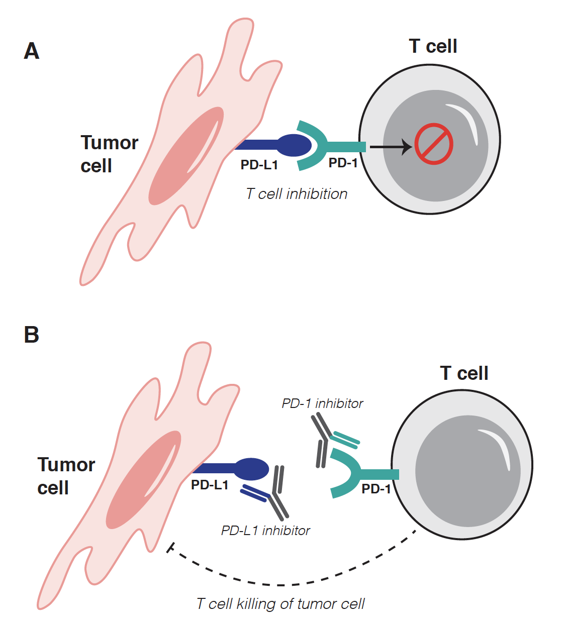
Figure 1. PD-1/PD-L1 interactions. A. Tumor cells upregulate PD-L1, which binds to PD-1 on T cells and suppresses their activity. B. PD-1 and PD-L1 checkpoint inhibitor drugs bind to the surface of these proteins, thereby blocking their interaction and preventing T cell suppression. T cells are then able to mount antitumor responses.