Flipping the Switch: The Hidden Roles of Class I Histone Deacetylases (HDAC’s)
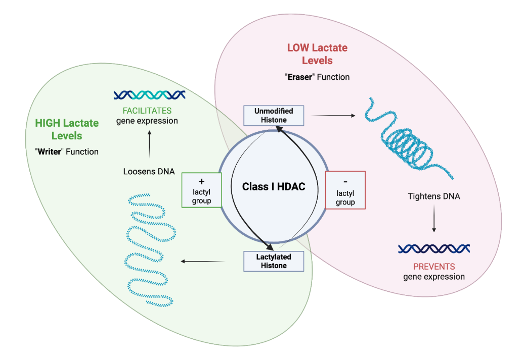
Figure 1: Demonstrates impacts of lactate levels on Class 1 HDAC activity. Under high lactate concentrations, lactate promotes the “writer” function that loosens DNA and facilitates gene expression. On the contrary, low lactate levels kickstarts the “eraser” function which tightens DNA and inhibits gene expression.
Written by Sid Pradeep, Schematic by Lexi Bean and Sid Pradeep
Background
Proteins are the main actors of cellular functions in our bodies. Adding particular molecules to proteins, sort of like accessorizing, can essentially switch them on or off. Once activated, they can serve a number of purposes - e.g. convert food to energy, enable defense against pathogens, enable cell-to-cell communication, and transport oxygen to name a few.
The process of activating proteins through alteration is known as post-translational modification (PTM), where proteins undergo changes after they are built by ribosomes (the “protein factories” of the body). While there are many different types of PTMs (over 650 and counting), the article “Class I Histone Deacetylases Catalyze Lysine Lactylation” by Drs. Gonzatti, Hinzten, Sharma, et. al (scientists both from UCSF as well as the University of Pennsylvania), specifically deals with a type of PTM called lysine lactylation (Kla), which is the addition of a molecule called lactate.
Previously, lactate was simply seen as a side effect - a toxic waste product - of glycolysis. Glycolysis is the breakdown of glucose to create energy for the body. This process happens all day long in every cell. However, scientists have recently started looking into the ways that lactate impacts the different genes from DNA and how they are expressed in the body.
In every cell, DNA is not just free-floating. Instead, it is wrapped by proteins called histones. These proteins control gene expression by binding to genes. When bound, they create extremely tight bonds that prevent molecules such as RNA polymerase and transcription factors from attaching to the gene and shutting off gene expression. On the contrary, when lactate is added to histones, the bonds with genes are loosened, thus, allowing for gene expression and interaction with the transcription factors.
What is this study about?
While Kla has mostly been associated with a specific type of lactate known as lactyl-coA (a lactyl group + an additional enzyme) that triggers Kla pathways by donating its lactyl group to proteins, researchers have found that there are numerous other molecules that have the potential to catalyze Kla - e.g., Class I histone deacetylases (HDAC’s 1, 2, and 3). In this study, Dr. Gonzatti, Hinzten, and Sharma along with their colleagues aimed to see the role of HDAC’s 1, 2, and 3 in Kla as well as compare them to a few other known Kla pathways. HDAC’s are an enzyme that are associated with removing lactyl groups from histone proteins, causing them to form tighter bonds with DNA. While there are numerous different types of HDAC’s, Class I HDAC’s are different from other forms of HDAC’s mainly because they are always found only in the nucleus of their target cells (while other forms of HDACs are able to move between the nucleus and cytoplasm). Previously, their role was only known to be involved in the reversal of Kla, which is the process of removing the lactyl group which tightens the DNA, stopping gene expression. However, researchers found that they are also useful for the forward reactions as well, and have the ability to lactylate histone proteins. Class 1 HDAC’s are unique in a few ways; in particular, they:
Are a cellular intermediate that’s independent of lactyl-coA dependent pathways and enables a different, simpler Kla process.
Controls the addition as well as the removal of lactyl chains, serving as both modifiers and de-modifiers.
Enables Kla reactions at normal levels of lactate unlike other metabolites that require unusually high concentrations. They are also reversible and the enzyme is able to switch its function based on the concentration of lactate levels.
To better understand the role of Class 1 HDAC’s, the research team conducted a series of experiments with the following results.
How was the study conducted?
The researchers organized a number of different experiments to test various components of Class 1 HDAC mediated Kla. They primarily looked at factors such as Class 1 HDAC co-activity with other key regulatory cells, comparison to other key Kla pathways, and effects of lactate levels on Class 1 HDAC activity. They also conducted further experiments to test the reversibility of Class 1 HDAC mediated Kla, which offers a significant breakthrough that expands the understanding of the function of these molecules.
What was discovered?
The results of the key experiments of this study are outlined below:
How do HDAC’s respond to infection?
This experiment aimed to see how Class 1 HDAC’s function with other key immune cells, specifically, macrophages, which are white blood cells that act as the body's first line of defense by “eating” invaders like bacteria. Researchers stimulated immune cells by releasing chemicals that under infection serve almost like a cellular “alarm clock” and macrophages were able to accelerate the secretion of lactate (by kickstarting glycolysis), causing a surge in Kla levels. When the macrophages were treated with Class 1 HDAC inhibitors, the accumulation of Kla was blocked. Since the cellular lactate levels were not changed, the authors suggest that macrophages require Class 1 HDAC’s to signal Kla.
2. Are HDAC’s what drive Kla or is it something else?
The next experiment aimed to see the comparative significance of Class 1 HDAC mediated Kla with respect to other key pathways known to catalyze Kla. They conducted experiments, isolating each pathway to monitor overall Kla levels. They found that none of these pathways other than Class 1 HDAC’s were required for Kla formation, showing how this is the primary mechanism for the Kla process.
3. HDAC’s are erasers of Kla, but are they effective to activate the process?
The researchers also tested the reversibility of Class 1 HDAC mediated Kla and found out that the same active site (area of the enzyme where the Kla reaction takes places) was required for inhibiting/promoting Kla, which supports the reversible role of the Class 1 HDACs. Essentially this proved how Class 1 HDAC’s serve as both modifiers and de-modifiers of Kla.
4. Does HDAC activity depend on lactate?
The final experiment was targeted towards testing the significance of lactate concentration by manipulating levels of Class 1 HDACs as well as lactate levels. In settings of high lactate availability, increasing the amount of Class 1 HDAC’s lead to higher Kla levels. In settings of low lactate, increasing the amount of Class 1 HDAC’s delactylated the proteins which shows how HDAC activity/role is dependent on lactate concentration.
Overall, these experiments highlighted not only the adaptive pathways of HDAC activity, but also their presence and interactions with other key cells, making them a crucial indicator of protein modification in the human body.
Why does it matter?
The research shows how the primary drivers of Kla levels under normal lactate conditions are Class 1 HDACs. This lays the foundation for further research into the role of these molecules, specifically, looking at the roles of Class 1 HDAC’s for crucial processes like cell-to-cell communication, generating energy for the cell, and sensing/locating nutrients in the body. Overall, this paper offers a scientific breakthrough on the role of Class 1 HDAC’s, as well as lactate in the human body, showcasing the extremely complex and diverse roles of cells that allow us to survive.
Reference:
Gonzatti MB, Hintzen JCJ, Sharma I, Najar MA, Tsusaka T, Marcinkiewicz MM, Da Silva Crispim CV, Snyder NW, Burslem GM, Goldberg EL. Class I histone deacetylases catalyze lysine lactylation. J Biol Chem. 2025 Oct;301(10):110602. doi: 10.1016/j.jbc.2025.110602. Epub 2025 Aug 18. PMID: 40835008; PMCID: PMC12624779.