Written by Arushi Chamaria, Schematics by Seha Kaur and Arushi Chamaria
Multiple sclerosis (MS) is a disease where the immune system attacks the brain and spinal cord, gradually damaging the wrapping around nerve fibers responsible for movement, sensation, and cognition. This ultimately reduces the speed and accuracy of nerves to communicate. As MS progresses, these structures shrink, (AKA atrophy), and the rate of this shrinkage can signal how quickly the disease is advancing. One of the most powerful tools doctors and researchers use to track disease progression is magnetic resonance imaging (MRI), which measures the volume of the spinal cord and brain over time. However, here's the problem: when patients first start taking medicines to help their MS, disease-modifying therapies (DMTs), the treatments can trigger an abrupt, dramatic-looking volume loss on MRI scans. Not because the nervous system is actually shrinking, but because the drugs are reducing inflammation, which had been falsely "inflating" tissue volume. This misleading signal is called pseudoatrophy, and it has complicated researchers' ability to accurately measure whether a treatment is actually protecting the brain and spinal cord.
A new study published in Annals of Neurology by Sacco and colleagues at UCSF and team asks whether a common anti-inflammatory treatment, high-dose glucocorticoids (HDGs), might actually prevent this false shrinkage from muddying the picture.
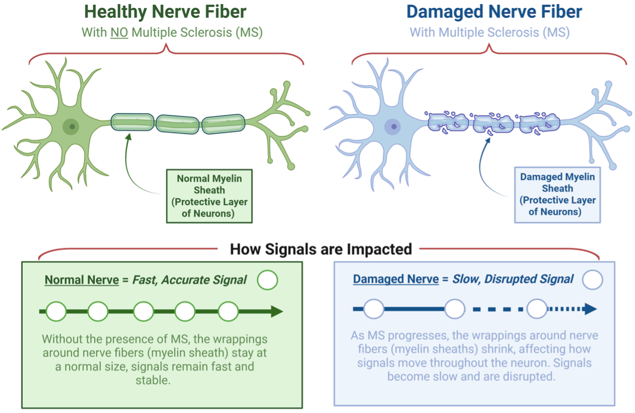
Figure 1: MS damages the myelin around nerve fibers, slowing signals. Left: healthy nerves send fast, accurate signals. Right: after MS drugs start, MRI can show a sharp drop in tissue volume that reflects inflammation and shrinkage (pseudoatrophy).
.
How Was It Conducted?
The researchers followed 123 newly diagnosed, treatment-naive (a person’s first exposure to treatment) MS patients from two existing UCSF cohort studies for up to three years. All participants had MRI scans of both the brain and the cervical spinal cord taken at baseline, and at two annual follow-up points. Of the 102 patients who began DMT treatment after their baseline scan, 42 had also received a course of HDGs in the roughly two months before that baseline scan, while 60 had not. A third group of 21 patients remained untreated throughout the first year and served as controls. Using sophisticated MRI analysis tools, the team tracked changes in the total cross-sectional area of the cervical spinal cord, further broken down into white matter and gray matter compartments, as well as several brain regions. Crucially, they compared first-year volume loss to second-year volume loss, since true pseudoatrophy should be front-loaded (biggest in year one, then tapering off), while real neurodegeneration would be more consistent over time.
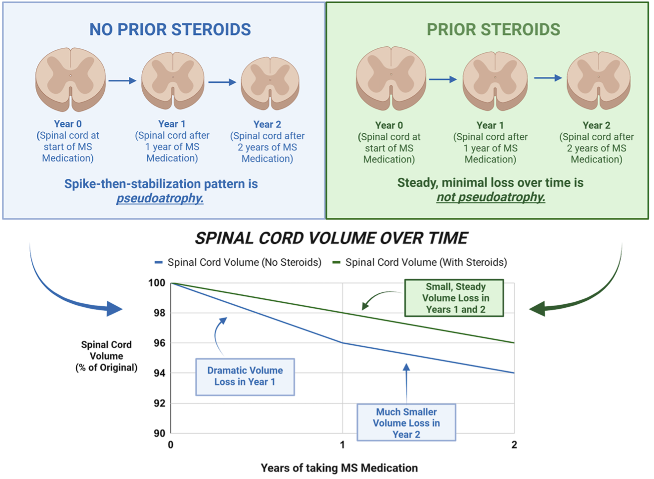
Figure 2: How the study tested whether steroids reduce this false signal. Left: untreated patients showed a big first-year volume drop that then leveled off (pseudoatrophy). Right: patients given steroids first showed steady, minimal loss (neurodegeneration).
The big picture: this could make clinical trials faster, cheaper, and more reliable.
Right now, MS drug trials often have to run for years just to see past the noise of pseudoatrophy in the first year. Some trials try to compensate by re-measuring patients at 6 months and using that as the "real" baseline, but that is a workaround with its own problems. If giving patients a short steroid course before a study begins can suppress the false signal entirely, trials could be shorter, need fewer participants, and produce clearer results. That means faster answers about whether new drugs actually work, and faster access to better treatments for people with MS.
What did they discover?
Patients who didn't receive steroids before starting treatment lost spinal cord volume four times faster in their first year than their second. It spiked dramatically right after treatment began, then slowed down. This is the hallmark of pseudoatrophy: a fake-out that makes it look like the nervous system is rapidly deteriorating when really the treatment is just reducing inflammation that had been quietly "puffing up" the tissue.
Patients who did receive steroids beforehand showed no such spike. Their rate of tissue loss was steady and low across both years. It is important to note that MS does cause real spinal cord atrophy over time, but in this short two-year window, the steroids had already reduced inflammation before the baseline scan, leaving nothing left to suddenly resolve when treatment started.
The spinal cord was hit three times harder by this effect than the brain. Even though pseudoatrophy has been studied in the brain for decades, this study showed the spinal cord is far more vulnerable to it. That's likely because the spinal cord is much smaller, so even a modest amount of inflammation takes up a proportionally larger share of its volume. When that inflammation clears, the drop looks enormous.
This effect only showed up in white matter, not gray matter, and that tells us something important about what's actually happening. White matter (the brain and spinal cord's "wiring") is directly affected by inflammation and blood-brain barrier leakage, so when steroids calm that down, white matter volume visibly drops. Gray matter (where the actual nerve cell bodies live) doesn't respond the same way to steroids, meaning its early volume loss may reflect real, ongoing damage rather than just inflammation clearing out. This distinction could help researchers separate "good shrinkage" (inflammation resolving) from "bad shrinkage" (actual nerve loss) for the first time.
Reference:
Sacco S, Papinutto N, Schoeps VA, Cheng S, Stern WA, Zhao H, Bischof A, Caverzasi E, Dombagahawatta M, Juwono J, Akula A, Cordano C, Beaudry-Richard A, Gomez R, Harms M, Santaniello A, Bove RM, Gelfand JM, Goodin DS, Green AJ, Oksenberg JR, Waubant E, Wilson MR, Zamvil SS, Cree BAC, Hauser SL, Henry RG. High-Dose Pulse Glucocorticoid Treatment Prevents White Matter Spinal Cord Pseudoatrophy in Newly Diagnosed Multiple Sclerosis. Ann Neurol. 2025 Oct;98(4):837-850. doi: 10.1002/ana.27298. Epub 2025 Jun 24. PMID: 40552528; PMCID: PMC12542327.