How Parasites Trigger Communication Between the Gut and Brain
Make it stand out
Whatever it is, the way you tell your story online can make all the difference.
Written by Zara Lee, Schematic by Lexi Bean & Zara Lee
Introduction
Have you ever noticed that when you’re sick, you suddenly don’t feel like eating or lose your appetite? Well, this doesn’t just occur as a side effect to sickness but is actually a coordinated response from the gut to your brain! Researchers knew that gut infections could influence the brain, but they didn’t know which cells started that process or how the message traveled from the intestine to the nervous system. In this study published in Nature, Touhara et al. from the David Julius lab worked with the labs of Yulong Li (Peking University, China), Stuart Brierley (South Australian Health and Medical Research Institute, Australia), and Richard Locksley (University of California, San Francisco) to investigate how parasitic infections trigger communication between specialized gut cells and the nervous system.
Background
As we know, the gut is more complex than just a digestive organ in our body. It contains millions of nerve cells and is consistently communicating with the immune system and the brain; often known as the gut-brain axis. This communication system not only helps with regulating digestion, but it also controls appetite and responses to sickness. The gut also acts as one of the body’s first warning systems as it detects harmful microbes and parasites before alerting the immune system and nearby nerves that something is wrong. The first line of defense in the gut lining are called epithelial cells. While the main job of these cells is to absorb nutrients, there are rare sensory epithelial cells that detect danger, including parasites. To understand this study, there are two types of sensory epithelial cells in the lining of the gut which are tuft cells and enterochromaffin cells (EC). Tuft cells help trigger immune responses and enterochromaffin cells (EC) release serotonin. When the EC cells receive a signal, they release serotonin, which makes up more than 90% of our body’s serotonin. Serotonin released by EC cells helps regulate gut movement, activates nearby nerve fibers, and supplies most of the serotonin moving throughout the body. Because both cell types become active during infection, scientists suspected that tuft cells and EC cells might work together to pass information from the intestine to the nervous system. Earlier studies by researchers including Richard Locksley and colleagues demonstrated that tuft cells respond to parasitic infections by releasing IL-25, helping stimulate a type 2 immune response. Other studies by David Julius’s group showed that EC cells release serotonin and communicate with sensory nerve fibers in the gut. Nevertheless, the scientists weren’t aware whether these two cell types directly interacted during infection or how certain interactions might influence behavior.
Study Method:
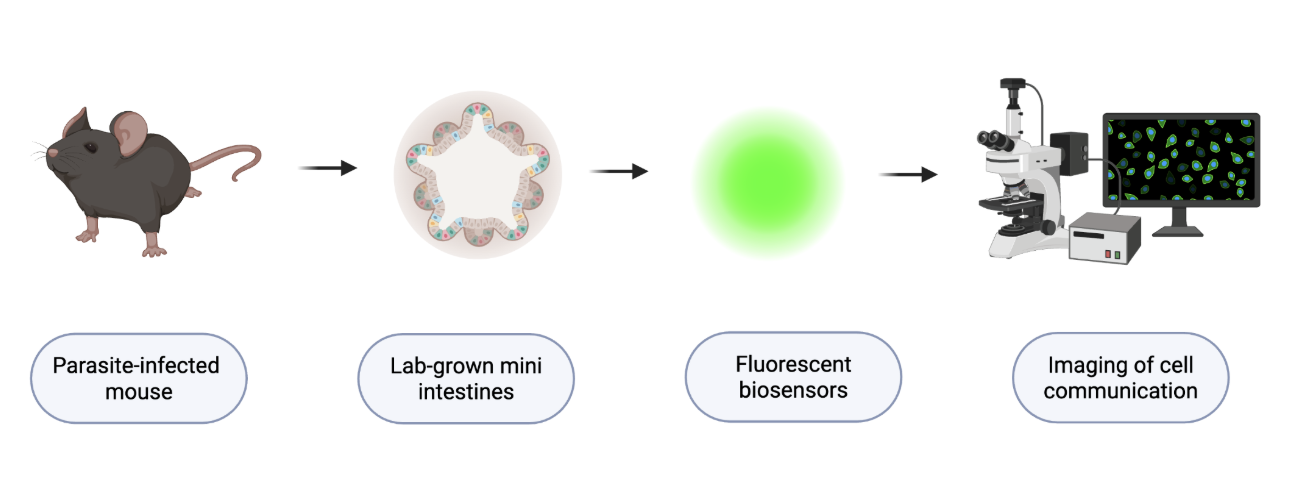
To investigate how the gut sends signals to the brain during a parasitic infection, Touhara et al., working in collaboration with the laboratories of Yulong Li at Peking University, Stuart Brierley at SAHMRI, and Richard Locksley at UCSF, studied mice infected with parasites that live in the intestine. They wanted to determine whether tuft cells could directly activate EC cells and whether that interaction was capable of alerting the vagus nerve, which carries messages from the intestine to the brain. To study this process, Touhara et al. used several mouse models, intestinal organoids (lab-grown mini versions of the intestine), biosensors with fluorescent light, and advanced imaging techniques that allowed them to watch signaling molecules being released by living cells in real time. Touhara and colleagues discovered that tuft cells release a signaling molecule called acetylcholine (ACh), serving as a messenger between cells. To their surprise, tuft cells were able to release acetylcholine in two different ways even though they lack the typical structure that nerve cells normally use for communication.
The first release of acetylcholine occurs immediately after parasites are found. Touhara et al. discovered that succinate (a molecule produced by specific parasites) provoked a quick burst of acetylcholine release from tuft cells, demonstrating that molecules released by parasites can activate tuft cells without first relying on signals from the immune system. The second release of acetylcholine occurred more slowly and was sustained over a longer period of time, usually occurring during the later stages of infection when the body has begun responding to the infection. To mimic the later stages of infection, Touhara and his collaborators exposed animals and organoids to immune signaling molecules called IL-4 and IL-25.
Both methods of acetylcholine signaling turn on or activate receptors on nearby EC cells. Yet, Touhara et al. found that only the sustained release of acetylcholine caused EC cells to produce enough serotonin to activate the vagus nerve. The vagus nerve is one of the body’s most important and major communication pathways between the gut and the brain. Once activated, these nerve fibers carry that message to the brain, suppressing appetite, which reduces food intake.
So, what did Dr. Touhara and colleagues ultimately discover?
The study by Touhara et al. revealed a two-step communication pathway that bridges parasitic infection to changes in behavior. Early in an infection, parasite-related molecules trigger a brief release of acetylcholine from tuft cells. As the infection progresses, a longer-lasting release of acetylcholine causes EC cells to produce enough serotonin to activate vagal nerve fibers that send signals to the brain. Together, these findings show how a parasite in the intestine can eventually influence the brain, explaining why an infection can cause someone to change behaviors in their appetite.
In this experiment, Touhara and colleagues monitored how much mice ate to see whether this pathway actually affects behavior. Mice with normal tuft-cell signaling ate much less during infection, in comparison with mice lacking tuft cells or acetylcholine signaling components, who ate normally. For the first time, Touhara et al. were able to trace back exactly on how an infection in the intestine leads to appetite loss. Instead of being a consequence of getting sick, eating less may help the body prioritize fighting off the infection and building back damaged tissue instead of using energy to digest food.
Why does it matter?
Before this study, researchers knew that parasites triggered immune responses and that EC cells could influence nearby nerves. However, no one had connected those two discoveries into one complete pathway until Touhara and his colleagues revealed it. They showed how tuft cells activate EC cells, which then stimulate the vagus nerve to relay information from the intestine all the way to the brain.
Because this pathway helps relay information from the gut and brain, it could eventually become a target for new treatments. Instead of broadly suppressing inflammation, future therapies might focus on specific signaling molecules, such as acetylcholine receptors or serotonin signaling, to restore normal gut-brain communication without negatively affecting the rest of the body. For example, individuals with inflammatory bowel disease (IBD) often experience chronic inflammation as well as appetite changes, nausea, and abnormal gut-brain signaling. Understanding how tuft cells, EC cells, and the vagus nerve normally communicate may help researchers understand why these symptoms develop and how they could eventually be treated more effectively.
Another surprising finding from this study is that many behaviors we associate with sickness aren’t just symptoms, but are responses. In this study, Touhara and his colleagues demonstrated that messages sent from the infected intestine can reduce appetite by activating the vagus nerve. Touhara et al. believed that eating less during infection may allow the body to redirect more energy toward fighting off the parasites and repairing the damaged tissue instead of digesting food. So, the next time you lose your appetite while being sick, remember that your intestine may be already sending signals through the vagus nerve to help your body recover faster.
Reference:
Touhara KK, Xu J, Castro J, Liang HE, Li G, Brizuela M, Harrington AM, Garcia-Caraballo S, O'Donnell T, Neumann D, Rossen ND, Deng F, Schober G, Li Y, Locksley RM, Brierley SM, Julius D. Parasites trigger epithelial cell crosstalk to drive gut-brain signalling. Nature. 2026 May;653(8114):465-473. doi: 10.1038/s41586-026-10281-5. Epub 2026 Mar 25. PMID: 41882357; PMCID: PMC13171452.